Billions of people are affected by chronic caries or tooth decay. Because there is no accurate measurement that predicts if tooth decay will occur at a specific location, there are no conventional means for managing this chronic disease. For most other common chronic diseases such as atherosclerosis, high blood pressure, diabetes, and cancers, the highest risk patients are identified by measuring leading indicators of disease and managing these indicators so that surgery is held for the last resort of treatment. Currently, tooth decay does not have a leading indicator for site-specific tooth decay that can be easily measured and tracked over time.
Multimodal laser-based angioscopy for structural, chemical and biological imaging of atherosclerosis
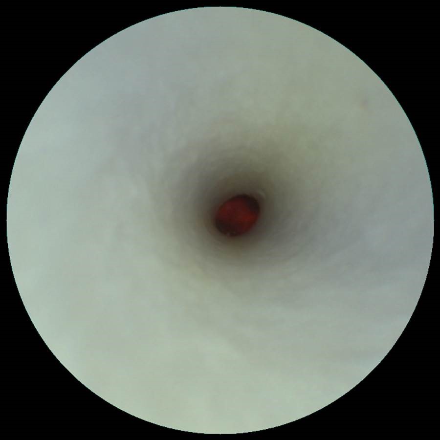
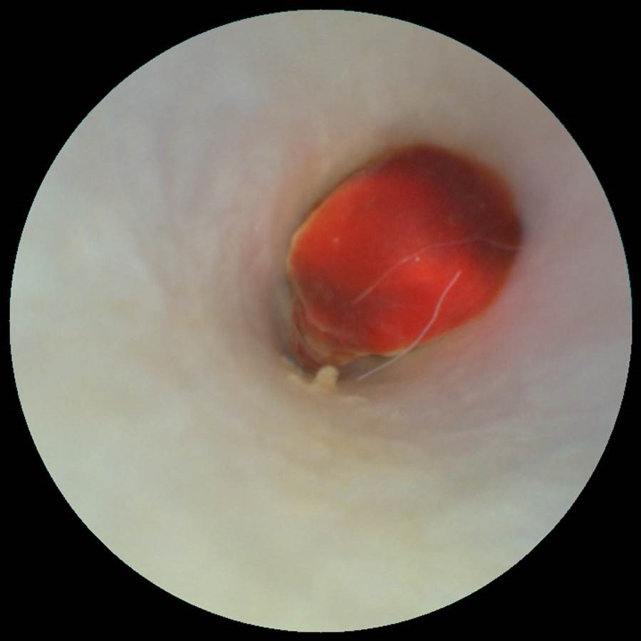
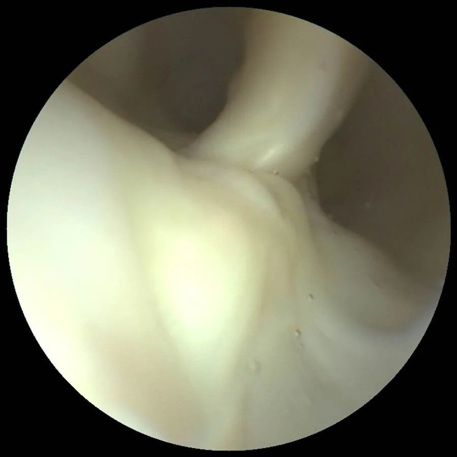
Multimodal SFE video acquisition of post-mortem arteries. Endoluminal endoscopic videos (Supplementary Videos 1,2,3,4,5) were obtained by navigating the SFE probe into the common carotid artery (CCA) toward the CB and then into the first 2 cm of the ICA and external carotid artery (ECA).
Researchers from the University of Michigan used a unique application of a medical camera to view the carotid artery to assess the risk of atherosclerosis. According to researchers at the University of Michigan School of Medicine used a scanning fiber endoscope, or SFE, to acquire high-quality images of potential atherosclerosis regions of the carotid artery that can be missed by conventional radiological techniques. "The camera actually goes inside the vessels," Dr. Luis Savastano is a Michigan Medicine resident neurosurgeon and first author of the study. He said in a press release, "We can see with very high resolution the surface of the vessels and any lesions, such as a ruptured plaque, that could cause a stroke. This technology may even be able to show which silent, but at-risk, plaques may cause a cardiovascular event in the future."