


Luce: Our First Product
Luce is a single-use, forward-viewing imaging catheter about one millimeter in diameter, with a console that shows a live color image of what lies directly ahead of the catheter, with digital zoom. VerAvanti intends to submit Luce for FDA 510(k) clearance for visualization in the peripheral and coronary vessels.
Our Unique Tech Features
The SFE platform is designed to deliver forward-looking, full-color imaging from a probe about one millimeter in diameter, at about 1.2 million pixels. Unlike side-viewing tools, it is intended to show what lies ahead of the catheter.


Imaging Modalities & Capabilities
Our SFE laser engine supports a growing range of imaging modalities, from standard color visualization to advanced spectroscopic and molecular techniques. Each modality runs through the same compact, single-fiber platform, expanding what is possible within the physical constraints of minimally invasive procedures.

True Color Videos
(Ruptured carotid atheroma with intraluminal clot.)
Defense & Aerospace Weekly

Microendoscopy
(Cholesterol deposits and villi in a gall bladder)
Defense & Aerospace Weekly

Molecular Imaging
(Fluorescent peptides specific for EGFR highlighting esophageal dysplasia)
Defense & Aerospace Weekly

Fluorescence Spectroscopy
(Carotid atheroma overlying elastin of tunica media)
Defense & Aerospace Weekly

Infrared pH sensing
(Dental Biofilm PH Heatmap)
Defense & Aerospace Weekly
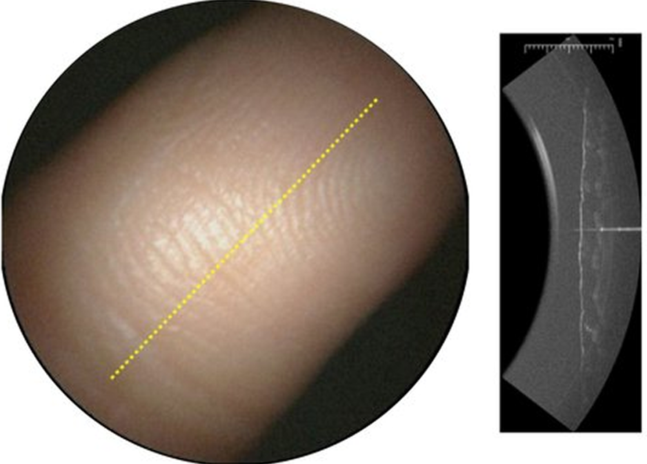
True-color + forward OCT
(Fingerprint)
Defense & Aerospace Weekly

Coherent Raman images
(Skin)
Defense & Aerospace Weekly

NIR multispectral images
(Tooth with dentin lesion)
Defense & Aerospace Weekly

Real-Time Forward Guidance
(Wire advancement toward an arterial occlusion —preclinical research image)
Defense & Aerospace Weekly

Targeted Biopsy Imaging
(Visualization of biological targets in high grade dysplasia in Barrett’s esophagus)
Defense & Aerospace Weekly
Potential Future Applications
VerAvanti's innovative imaging technology aims to extend its capabilities far beyond intravascular health, addressing critical needs across multiple medical fields. From gastroenterology and pulmonology to urology and dental care, our advanced solutions could empower clinicians with unparalleled diagnostic precision and therapeutic guidance.


A Look Inside Our Scanning Fiber Endoscope (SFE)


SFE generates images by projecting red, green, and blue laser light in a controlled spiral scanning pattern.
As the beam scans forward, reflected light is collected to reconstruct a full-color image in real time. This pixel-by-pixel acquisition enables continuous visualization with high sensitivity and efficient light use.

At the core of SFE is a piezo-actuated oscillator that drives the scanning motion of the optical fiber.
This mechanism is integrated within a probe measuring approximately 1 mm in diameter and a few millimeters in length, enabling precise beam control within an ultra-compact form factor.